Welcome to PICU Doc On Call, A Podcast Dedicated to Current and Aspiring Intensivists.
I’m Pradip Kamat coming to you from Children’s Healthcare of Atlanta/Emory University School of Medicine and I’m Rahul Damania from Cleveland Clinic Children’s Hospital. We are two Pediatric ICU physicians passionate about all things MED-ED in the PICU. PICU Doc on Call focuses on interesting PICU cases & management in the acute care pediatric setting so let’s get into our episode.
Here’s the case of a 12-week-old girl old who is limp and seizing presented by Rahul.
- Chief Complaint: A 12-week-old previously healthy female infant was found limp in her crib and developed generalized tonic-clonic seizures on the way to the hospital.
- History of Present Illness: The mother returned from work on a Saturday to find her daughter unresponsive in her crib. The infant had been left in the care of her mother’s boyfriend, who stated that the daughter had been sleeping all day and had a small spit up. As the patient continued to have low appetite throughout the day and continued to be unresponsive in her crib, mother called EMS to bring her to the emergency department. En route, the patient had tonic movement that did not resolve with intranasal benzodiazepines.
- ED Course: The infant presents to the ED being masked. Upon arrival at the ED, the infant was in respiratory distress, with a heart rate of 190 beats per minute, respiratory rate of 50 breaths per minute, and oxygen saturation of 85% with bagging. She was intubated for seizure control upon arrival at the ED. Physical examination in the ED revealed bruising on the right neck region but was otherwise unremarkable. A non-contrast head CT showed no acute intracranial abnormalities. The initial diagnostic workup revealed normal CBC, mildly elevated hepatic enzymes, and pancreatic enzymes which were within normal limits. The blood gas showed metabolic acidemia with PCO2 in the 60s.
- Admission to PICU: Upon admission to the PICU, neurosurgery and trauma teams were consulted. A skeletal survey and ophthalmology consult for a fundoscopic examination were ordered, as there were concerns of non-accidental trauma. Further investigation is underway to determine the cause of the infant’s condition.
To summarize key elements from this case, this patient has:
- Patient left with mother’s boyfriend
- Infant found limp and had seizures requiring intubation
- Neck bruise
- All of these bring up a concern for Non-Accidental Trauma (NAT) the topic of our discussion.
Let’s start with a short multiple-choice question:
Which imaging modality is the most appropriate for establishing a diagnosis of abusive head trauma (AHT) in a 12-week-old infant with an open fontanelle on the exam?
- A. CT scan of the brain without contrast B. MRI of the brain without contrast C. Skull X-ray D. Doppler ultrasound of the head
Rahul, the correct answer is A.
Though ultrasound may be less invasive, the penumbra effect in cranial ultrasound makes it hard to visualize the parts of the brain located just under the convexity of the skull such as a subdural hematoma. Regardless of the small radiation risk, noncontrast head CT is the method of first choice in imaging traumatic brain injury for both fractures and intracranial pathology. CT scan has a short scan time and is widely available. Non-contrast-enhanced CT has a high sensitivity for detecting acute hemorrhage and midline shift.
Thanks for that detailed explanation, I agree CT scan is a valuable diagnostic tool that provides detailed recon images for understanding the mechanism of fractures.
What about the role of MRI in diagnosing abusive head trauma?
- MRI has lower sensitivity for acute hemorrhage compared to a CT scan and takes longer to acquire images, which may require anesthesia to provide immobility. However, a systematic review by Kemp and colleagues published in 2009 (Clin Radiol. 2009;64:473–483) reported that MRI performed following an abnormal CT scan in children with abusive head trauma revealed new information in at least 25% of cases, such as cranial shearing, ischemia, infarction, parenchymal hemorrhages, and cerebral contusions. It’s important to note that the role of MRI in cases where the initial CT scan is normal is unclear. Additionally, MRI is more accurate in evaluating time points in certain lesions, making it a valuable tool in the diagnosis and management of abusive head trauma in pediatric patients.
💡 In summary, a CT scan is the preferred imaging modality for assessing traumatic brain injury in cases of suspected abusive head trauma, while cranial ultrasonography may be useful in some cases. It’s important to remember that interpretation of imaging in cases of suspected AHT requires complete clinical information.
Alright, Pradip, very interesting that our initial CT scan did not show any signs of bleeding, once the patient became more stable in the PICU, what did the skeletal survey show?
- The skeletal survey showed multiple fractures of varying ages, including multiple rib fractures, and an unhealed clavicle fracture. The team closely monitored the infant’s condition and initiated treatment as necessary.
Rahul, can you give us a brief introduction to non-accidental trauma in the pediatric ICU?
- Child abuse, also known as battered child syndrome, can take multiple forms such as physical abuse, sexual abuse, neglect, psychological maltreatment, general neglect, and medical neglect. Today, we’ll focus on physical abuse that intensivists may encounter in their practice.
- In the Pediatric Intensive Care Unit (PICU), the team is more likely to see cases of abusive head trauma, abdominal trauma, burns, complex fractures, and rib fractures, which may be identified when a chest radiograph is obtained after intubation. These are serious and often life-threatening conditions that require a multidisciplinary team approach and specialized care.
💡 To summarize, physical abuse in children, particularly infants, can present with nonspecific symptoms and signs, such as vomiting or apnea. This highlights the importance of considering the possibility of abusive head trauma in such cases.
Please also remember that the term, abusive head trauma replaced “shaken baby syndrome,” and it’s a serious and often life-threatening condition that requires prompt recognition and intervention. Therefore, it’s essential for us as intensivists to be familiar with the various forms of physical abuse, including abusive head trauma, and work closely with other specialists to ensure that the patient receives the best possible care.
Pradip, let’s dive deep into abusive head trauma, do you mind talking about the spectrum of symptoms we can see?
Abusive head trauma is the most common presentation of child abuse in the PICU: As seen in our case presentation infants may present with apnea, altered mental status, loss of consciousness, limpness, vomiting, seizure, poor feeding, or have subtle signs like swelling of the scalp.
In a third of abusive head trauma cases, the infant was seen by another physician in the preceding 2-3 weeks. The diagnosis requires a high level of suspicion especially in an infant with fractures, ecchymosis, and failure to gain weight. AHT is the leading cause of fatal injuries in children.
📖 AHT is responsible for 53% of all severe TBI cases in infants.
What is the pathophysiology of injury in abusive head trauma?
The pathophysiology of abusive head trauma in infants is complex and multifactorial. The skull of a neonate is soft and malleable, which allows forces applied to the skull to propagate directly to the brain tissue. Additionally, the higher water content and lack of myelination make the brain more susceptible to shearing forces, which occur with shaking. Infants have a larger head in proportion to their body, constituting about 15-20% of total body weight as opposed to 2-3% in adults.
So, we’ve discussed how the pathophysiology of abusive head trauma in infants is complex and multifactorial. Can you tell me more about how the soft and malleable skull of a neonate plays a role in this type of injury?
- A heavier head with a lack of nuchal muscular strength predisposes the head to sustain severe injury as opposed to an older child. Furthermore, due to a lack of coordination of the head and body motion, the infant is unable to protect themselves. Injuries in abusive head trauma can be due to blunt impact, shaking with blunt impact, or shaking alone. Whiplash shaking and jerking subjects the brain to rotational acceleration and deceleration forces, which explains brain injuries and retinal hemorrhages in the absence of external trauma. The resulting traumatic brain injuries can have devastating and long-lasting effects on the child’s cognitive and physical development.
Rahul, how would an intensivist assess a child with physical abuse?
- As the pediatric intensive care unit is a team sport, it’s important to consult with multiple teams early on in cases of suspected abusive head trauma. This includes the trauma and neurosurgery teams, radiologists, child advocacy services, and social workers. In some states, early referral to Child Protective Services or law enforcement is mandatory to protect other siblings from harm. By involving these specialized teams and agencies, we can ensure a comprehensive approach to the diagnosis and management of abusive head trauma in pediatric patients.
- Absolutely, Rahul. The first step in diagnosing abusive head trauma is to obtain a detailed history from parents or caregivers. It’s important to determine if the child was brought for medical attention or neglected after the traumatic event. Additionally, we need to assess whether the child’s development level is consistent with the proposed mechanism of injury and whether the alleged events account for all injuries.
What are some key historical features that can help diagnose child abuse in cases of suspected abusive head trauma?
- In a retrospective study of 163 children, 30% of whom met the criteria for physical abuse, certain historical features had high specificity and positive predictive value for diagnosing child abuse. Having no history of trauma had a specificity of 0.97 and a positive predictive value of 0.92 for abuse. Among the subgroup of patients with persistent neurological abnormality at hospital discharge, having a history of no or low-impact trauma had a specificity and positive predictive value of 1.0 for definite abuse.
A detailed history is crucial in diagnosing abusive head trauma, as certain negative historical features such as no history of trauma and low-impact trauma have high specificity and positive predictive value for diagnosing child abuse when the clinical suspicion is high
- Certainly. In our case, the mother’s boyfriend claimed that the baby fell from the crib onto the hardwood floor. However, falls from less than five feet are unlikely to cause moderate or large subdural hematomas in children and are rarely fatal. It’s important to note that scalp contusions or lacerations are common in such falls, while a skull fracture is typically linear and located in the parietal region without associated intracranial hemorrhage.
- Rahul, in our case the patient had mild transaminitis, can you comment on abusive abdominal trauma?
- Certainly, abdominal trauma in the PICU is an important topic to discuss. In our case, the patient had mild transaminitis which leads us to question the possibility of abusive abdominal trauma. It’s important to note that AAT is actually the most common cause of abdominal injuries in children under two years of age.
- The outcome for patients with AAT is also worse than those with accidental trauma, with a mortality rate ranging from 9-30%, as opposed to 4.7% for those with accidental injuries. Symptoms such as vomiting may be initially attributed to medical conditions like gastroenteritis, which can lead to a delay in diagnosis. The most common injuries in AAT involve the liver, kidney, spleen (with the liver being more common than the spleen), and the stomach/intestines. If a child presents with pancreatitis after a “reported fall,” it should raise suspicion for abusive abdominal trauma.
Let’s keep building on this diagnostic framework, besides history what else would you emphasize?
- Certainly, in addition to obtaining a thorough history, the next step in evaluating a child for non-accidental trauma in the PICU is to conduct a comprehensive physical exam. It’s essential to document any skin findings, oral lesions, or eye findings, as well as to take photographs and place them in the patient’s electronic medical records with the appropriate date/time. The next step is to obtain imaging, with CT being most helpful in the acute phase to determine the need for neurosurgical intervention, while MRI may be needed to evaluate for diffuse axonal injury, ischemia, cranial shearing, or infarction.
- A skeletal survey should also be obtained to assess for fractures, and if abdominal injuries are suspected, a CT or MRI of the abdomen should be obtained. Additionally, CBC, CMP, coagulation studies, and pancreatic enzymes should be ordered. An ophthalmology consult for retinal hemorrhages is crucial, as they cannot be specifically dated and may clear quickly, so early examination is important. Lastly, postmortem examination is recommended for children who died from unexplained causes or abusive injuries.
To summarize, retinal hemorrhages are a common finding in fatal cases of AHT seen in 85% of cases with a spectrum of disease such as extensive hemorrhages leading to retinal tears, detachment, and vitreal hemorrhage. While retinal hemorrhages are not specific to AHT, they can be easily distinguished based on history, imaging, and clinical evaluation. Conditions such as birth trauma can cause retinal hemorrhages; the presence of these retinal hemorrhages can be correlated with the mode of delivery, with vacuum extractions having a higher correlation compared to NSVD and C-sections. It is important to note that retinal hemorrhages should not be attributed to birth trauma after 6 weeks of age. Other differentials for retinal hemorrhages in infants to keep in mind include leukemia, meningitis, vasculitis, and severe hypertension. However, by and large, please keep NAT on top of your differential.
How would you outline your general management framework if the history, physical examination, and diagnostic investigation suggest a diagnosis of abusive head trauma?
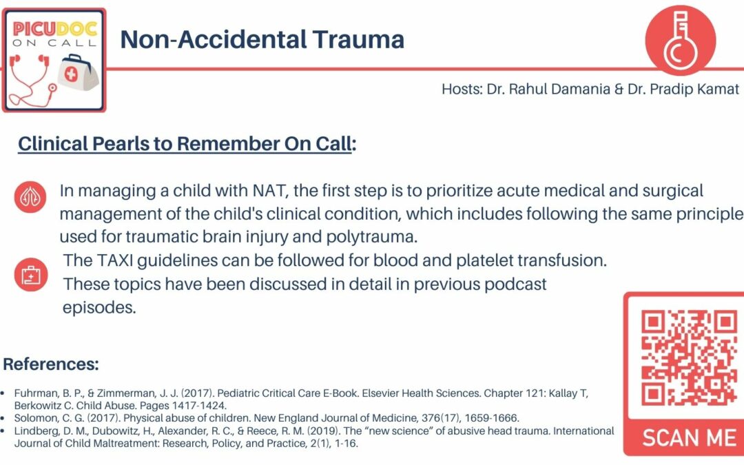
- In managing a child with NAT, the first step is to prioritize acute medical and surgical management of the child’s clinical condition, which includes following the same principles used for traumatic brain injury and polytrauma. This involves early consultation with neurosurgery and trauma teams, implementing cerebroprotective measures for intracranial pressure management and prevention of secondary brain injury, using lung protective ventilation strategies, providing adequate analgosedation, maintaining judicious fluid balance, and correcting any necessary laboratory abnormalities. The TAXI guidelines can be followed for blood and platelet transfusion. These topics have been discussed in detail in previous podcast episodes.
Rahul, let’s close this episode with some key summary take-homes.
Our case highlighted the importance of maintaining a high index of suspicion for non-accidental trauma in infants and young children. The infant in our case had clinical findings inconsistent with the history provided by the caregiver, leading to a diagnosis of abusive head trauma. Abusive abdominal trauma should also be considered in cases of non-accidental trauma, with a high mortality rate and common injuries to the liver, kidney, spleen, and intestines. A team approach is crucial in the management of NAT in the PICU, involving specialists from trauma, neurosurgery, child advocacy, radiology, and social services. Early recognition and intervention are essential in improving outcomes for these vulnerable patients.
This concludes our episode on child abuse We hope you found value in our short, case-based podcast. We welcome you to share your feedback, subscribe & place a review on our podcast! Please visit our website picudoconcall.org which showcases our episodes as well as our Doc on Call management cards. PICU Doc on Call is co-hosted by myself Dr. Pradip Kamat and Dr. Rahul Damania. Stay tuned for our next episode! Thank you!
- References
- Fuhrman, B. P., & Zimmerman, J. J. (2017). Pediatric Critical Care E-Book. Elsevier Health Sciences. Chapter 121: Kallay T, Berkowitz C. Child Abuse. Pages 1417-1424.
- Solomon, C. G. (2017). Physical abuse of children. New England Journal of Medicine, 376(17), 1659-1666.
- Lindberg, D. M., Dubowitz, H., Alexander, R. C., & Reece, R. M. (2019). The “new science” of abusive head trauma. International Journal of Child Maltreatment: Research, Policy, and Practice, 2(1), 1-16.

