Welcome to PICU Doc On Call, A Podcast Dedicated to Current and Aspiring Intensivists.
I’m Pradip Kamat and I’m Rahul Damania. We are coming to you from Children’s Healthcare of Atlanta – Emory University School of Medicine.
Welcome to our Episode an 8-year-old admitted for PRESS syndrome with altered mental status secondary to seizures.
Here’s the case presented by Rahul:
Our patient today is an eight-year-old who was admitted to the floor with a diagnosis of MIS-C. On his initial echo, his EF had mildly depressed systolic function, dilatation of coronaries, and worsening of inflammatory markers. As a result, the care team increased the dosing of the methylprednisolone administered to this patient. Since the initiation of methylprednisone, The patient’s SBP had been steadily increasing with the latest systolic values approaching 140s-150s.
On hospital day 3 patient had a generalized tonic-clonic seizure and became unresponsive for which a rapid response on the floor was called. The patient was emergently bagged and brought to the PICU for airway protection and intubation
Initial vitals on PICU admission: He was afebrile, mildly tachycardic, and hypertensive to 160s even after sedation.
In the PICU an initial head CT scan done after intubation and stabilization of the patient showed no bleeding or mass. cEEG monitoring was initiated, neurology consulted and an MRI was ordered for the following day. As his AMS was thought to be related to his BP, the team pursued BP control with Nicardipine.
To summarize key elements from this case, this patient has:
- Seizure
- Altered mental status
- Hypertension
- Acute respiratory failure
- All of which brings up a concern for an acute CNS pathology.
Absolutely, the differential is broad, however, right now I am thinking of an acute stroke categorized as hemorrhagic, ischemic, or venous thrombotic; a meningoencephalitis, CNS vasculitis, acute demyelinating encephalomyelitis, metabolic encephalopathy, tumor, or AMS related to hypertension.
Pradip, let’s transition into some history and physical exam components of this case?
What are key history features in this child?
- MIS-C with cardiac dysfunction and coronary anomalies
- Increase in steroid dosage
- Progressive increase in BP as a result of this increase
Rahul, are there some red-flag symptoms or physical exam components which you could highlight?
- The patient’s physical exam was relatively normal. Of note, the fundoscopic exam did not reveal papilledema and no renal bruit was auscultated.
- His Pupils were equal, round, and reactive to light. The face was symmetric. Normal bulk and tone. The patient was sedated and did not withdraw extremities to noxious stimuli. Tendon reflexes were equal throughout. and no clonus is noted. Fundoscopic exam revealed no papilledema which may rule out increased ICP as a cause for our AMS.
To continue with our case, Rahul ,what were the patient’s labs were consistent with:
- Down trending CRP, ESR, BNP, and troponin
- ECHO is consistent with improved cardiac function as well as improvement of coronary dilatation.
- CT scan with no bleed
- MRI suggestive of changes in the posterior brain with distinct edema pattern
OK to summarize, we have:
An eight-year-old, with acute severe hypertension, seizure altered mental status, and MRI changes suggestive of vasogenic edema in the posterior part of the brain -all this brings up the concern for posterior reversible encephalopathy syndrome (PRES) the topic of our discussion today.
Rahul ,Let’s start with a short multiple-choice question:
A 19-year-old with h/o of renal transplant on tacrolimus and recent initiation of steroids for rejection presents with acute severe hypertension and a GTC seizure. The patient is afebrile with no rash. CT scan at OSH reveals no mass or hemorrhage. After stabilization and initiation of antihypertensive therapy, the next study of choice for diagnosis is
- A) Continuous EEG
- B) MRI
- C) Lumbar puncture
- D) Positron emission test (PET scan) of the brain
Rahul, the correct answer is B) MRI. Patients such as the one described in the above question are at high risk to develop PRES. MRI will show classic changes associated with PRES- Involvement of the parieto-occipital region of the brain. Vasogenic edema (typically affecting the brain white matter) is characterized by hyperintensity on FLAIR and T2-weighted MRI sequences. As seizure is a presentation of PRES as in our case above, cEEG monitoring especially if intubated is indicated but may not be helpful in diagnosis. An LP also will not help with the diagnosis of PRES and the patient in this question is afebrile. PET scan may have a role in unusual or atypical cases of PRES mainly to distinguish it from the tumor. There is decreased fluorodeoxyglucose (FDG) and Methionine(MET) uptake in most PRES cases compared to tumors such as gliomas or lymphomas.
To summarize:
The diagnosis of PRES relies on a combination of clinical presentation and neuroimaging. Acute or subacute presentation with encephalopathy, generalized tonic-clonic seizures (60-75% patients), headaches, visual field deficits, cortical blindness, hallucinations, or rarely focal findings such as aphasia or hemiparesis should raise suspicion for PRES. Headache+visual disturbances+generalized tonic-clonic seizures =PRES unless proven otherwise.
Rahul, as you think about our case, what would be your differential?
- Infectious encephalitis (CSF is abnormal, CSF gram stain, CX or PCR)
- CNS vasculitis (CSF pleocytosis, cytotoxic edema in a non-PRES like pattern)
- Acute demyelinating encephalomyelitis (ADEM): H/o URI/bacterial infection, fever, usually asymmetric involvement of supratentorial regions on imaging
- malignancy or tumor (glioma or lymphoma)-Typically subacute-chronic presentation, h/o malignancy, absence of quick resolution, abnormal CSF or blood work
- Another important grouping I would consider is a toxidrome — some of these we mention in our prior podcast episodes so listeners please check them out!
Rahul, can you comment on the pathogenesis of PRES**
It is hypothesized that when the patient’s mean arterial BP exceeds the upper limits of cerebral autoregulation it leads to hyper-perfusion and the breakdown of the blood-brain barrier allowing interstitial extravasation of plasma and macromolecules and subsequently vasogenic edema.
- PRES can also develop in patients (15-20%) with normal BP or hypotension, which does not exceed the auto-regulatory capacity of the cerebral blood flow.
- In these patients, the endothelial dysfunction and breakdown of the blood-brain barrier could be from the cytokines and inflammatory mediators from systemic toxic effects of medications, etc. resulting in vasogenic edema. The posterior regions of the brain are more susceptible to vasogenic edema because little sympathetic innervation exists in the posterior fossa.
Pradip, which patients are at risk for PRES?
Apart from acute severe hypertension, a number of other conditions are associated with PRES. PRES is seen in patients receiving immune suppression, especially calcineurin inhibitors (tacrolimus or cyclosporine) after a stem cell or solid organ transplantation. Higher incidence is seen in BM or stem cell transplant as the dose of immunosuppression is higher in these patients compared to solid organ transplant patients. Autoimmune disorders, pregnancy with pre-eclampsia, eclampsia as well as those with renal disease have been linked to PRES.
Here’s a summary point when you see progressive hypertension in a patient post-transplant, after doing due diligence to pain control and diagnostic workup, pay close attention to mental status as these patients (especially if they are immunosuppression) are at high risk for developing press.
Pradip If you had to work up this patient, what would be your diagnostic approach?
- Acute-subacute symptoms such as confusion, seizures, visual, headache, and visual symptoms in the presence of hypertension, immune suppression, auto-immune disorders, transplant, or pre-eclampsia are highly suggestive of PRES.
- Brain imaging is the most important diagnostic test typically used to exclude other diagnoses. The presence of vasogenic edema affecting the white matter in the parieto-occipital regions of both hemispheres (rarely asymmetric) on MRI FLAIR is highly sensitive. Although vasogenic edema can be seen in other areas of the brain there is always concomitant involvement of the parieto-occipital regions. Some patients with PRES may have restricted diffusion (15-30%) seen in larger areas of vasogenic edema, some patients (10-25%) may have intracranial or subarachnoid hemorrhage, especially in allogeneic BMT patients.
- Other investigations which need to be decided on a case-by-case basis include obtaining LP, looking for CSF pleocytosis, gram-stain/Cx, PCR for viruses as well as cytology for malignancy.
- Baseline blood gas, CBC, CMP, DIC may be required depending on the severity of the presentation
- EEG: As 60-75% of patients with PRES can present with generalized tonic-clonic seizures and PRES can be suspected as the underlying cause of status epilepticus (especially when bilateral occipital sharp waves are present) continuous EEG monitoring especially in an intubated patient must be initiated.
The presence of vasogenic edema affecting white matter in the parieto-occipital regions of both hemispheres (rarely asymmetric) on MRI FLAIR in the appropriate clinical context is highly sensitive for PRES. Neuroimaging helps to exclude alternative diagnoses such as brain tumor or acute demyelinating syndromes as well as detection of intracranial hemorrhage.
Rahul, if our history, physical, and diagnostic investigation led us to PRES as our diagnosis what would be your general management of framework?
- There is no specific management for PRES.
- Treatment is supportive and basic principles of good ICU care are paramount for optimal outcomes.
- Any treatable underlying cause such as severe hypertension must be managed appropriately. The anti-epileptic agent should be initiated and may be required for 3-12months (Morris EB et al. Pediatr Blood Cancer 2007). In our case, the patient’s high BP was triggered by high-dose steroids used for MIS-C. The steroid dose was reduced as the patient’s BP was controlled using IV nicardipine. A collaborative approach with rheumatology, infectious diseases, and ICU helped make decisions that allowed lowering of the steroid dose and titrating the BP. Magnesium has been used in PRES due to pre-eclampsia and eclampsia for seizure prophylaxis/treatment. The management of PRES from induction chemotherapy is difficult as deciding which meds to halt in a patient receiving multiple agents may not be easy. If PRES is caused by anti-rejection meds in a transplant patient, that medication may need to be discontinued at least temporarily. Listeners are encouraged to listen to our podcast on acute severe hypertension on how to safely lower BP in patients with acute severe hypertension.
- Can you also comment on the prognosis of PRES?
- Overall the prognosis of PRES is excellent although some severe forms may not be fully reversible and can have a mortality rate of 3-6%. Mortality is due to intracranial bleeding, cerebral edema in posterior fossa, hydrocephalous or diffuse cerebral edema, or raised ICP. In 10-20% of patients with PRES there can be neurological sequelae in the form of seizures, neuro-deficits such as hemiparesis, decreased visual activity, and dizziness. In 5-10% of patients PRES can be recurrent especially in patients with difficult to treat hypertension or who are on medications for their organ transplant.
- Key objective take-aways from today’s episodes:
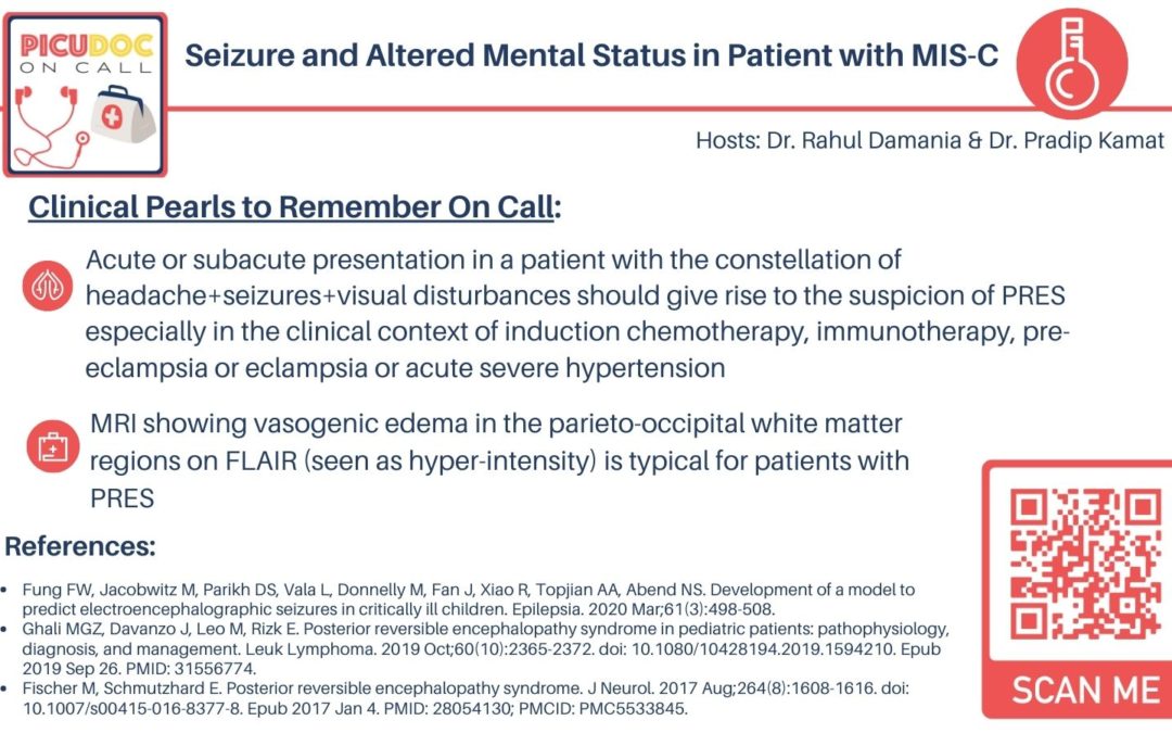
- Acute or subacute presentation in a patient with the constellation of headache+seizures+visual disturbances should give rise to the suspicion of PRES especially in the clinical context of induction chemotherapy, immunotherapy, preeclampsia or eclampsia or acute severe hypertension
- MRI showing vasogenic edema in the parieto-occipital white matter regions on FLAIR (seen as hyperintensity) is typical for patients with PRES
This concludes our episode on posterior reversible encephalopathy (PRES) We hope you found value in our short, case-based podcast. We welcome you to share your feedback, subscribe & place a review on our podcast! Please visit our website picudoconcall.org which showcases our episodes as well as our Doc on Call management cards. PICU Doc on Call is hosted by me Pradip Kamat and my co-host Dr. Rahul Damania. Stay tuned for our next episode! Thank you!
References
- Fugate JE, Rabinstein AA. Posterior reversible encephalopathy syndrome: clinical and radiological manifestations, pathophysiology, and outstanding questions. Lancet Neurol. 2015 Sep;14(9):914-925. doi: 10.1016/S1474-4422(15)00111-8. Epub 2015 Jul 13. Erratum in: Lancet Neurol. 2015 Sep;14(9):874. PMID: 26184985.
- Ghali MGZ, Davanzo J, Leo M, Rizk E. Posterior reversible encephalopathy syndrome in pediatric patients: pathophysiology, diagnosis, and management. Leuk Lymphoma. 2019 Oct;60(10):2365-2372. doi: 10.1080/10428194.2019.1594210. Epub 2019 Sep 26. PMID: 31556774.
- Fischer M, Schmutzhard E. Posterior reversible encephalopathy syndrome. J Neurol. 2017 Aug;264(8):1608-1616. doi: 10.1007/s00415-016-8377-8. Epub 2017 Jan 4. PMID: 28054130; PMCID: PMC5533845.
More information can be found
- Fuhrman & Zimmerman – Textbook of Pediatric Critical Care Chapter 61 (page 749-750); Chapter 64 (page 781); Chapter 78 (page 951); Chapter 92 (page 1111)
- Key objective takeaways:
- Acute or subacute presentation in a patient with the constellation of headache+seizures+visual disturbances should give rise to the suspicion of PRES especially in the clinical context of induction chemotherapy, immunotherapy, preeclampsia or eclampsia or acute severe hypertension
- MRI showing vasogenic edema in the parieto-occipital white matter regions on FLAIR (seen as hyper-intensity) is typical for patients with PRES

