Welcome to PICU Doc On Call, A Podcast Dedicated to Current and Aspiring Intensivists.
I’m Pradip Kamat and I’m Rahul Damania and we are coming to you from Children’s Healthcare of Atlanta – Emory University School of Medicine.
Welcome to our episode of a 14-year-old girl with sudden acute outbursts of aggression and severe agitation.
Here’s the case presented by Dr. Damania:
A 14-year-old previously healthy teenager with no significant past h/o presents to the PICU with a three-day h/o of aggressive behavior, agitation, and screaming. Her mother reports that her daughter has recently developed insomnia, abnormal movements and is more irritable with temper tantrums and episodic unintelligible verbal output. Parents report no recent stressors at home or at school. She has been also complaining of headaches for the past week along with things “being too loud”. She denies any vertigo symptoms or tinnitus. The patient is brought to the ER due to persistent auditory/visual hallucinations followed by agitation, aggressive behavior, and catatonia. There is no h/o of recent illnesses, head trauma, fevers, rash, abdominal pain, diarrhea, or vomiting. Social history is negative for drugs of abuse in the home. Family h/o negative for seizures, and psychiatric disorders.
The patient is sent to the ED and upon arrival has an unprovoked convulsive episode concerning a GTC seizure. The patient was initially admitted to the floor but transferred to the PICU for management of severe agitation, aggressive behavior, and fluctuations of blood pressure and heart rate.
Initial vitals in the PICU were notable for tachycardia. The patient was found to be afebrile, normotensive for age, and SpO2 96% on RA. Her physical exam though limited by her aggressive behaviors was normal. The heart, lung, and abdominal exams are normal with no rash or bruising on her body.
Initials lab work includes a negative:
- U preg
- Serum and Urine tox screen
- CBC, CMP, and UA are all within normal limits
- Inflammatory markers — including ESR CRP are unremarkable.
- A head CT which was normal and an A lumbar puncture revealed colorless CSF with 8 white and 0 red cells. Serum and CSF glucose were within normal limits and protein count in CSF was negligible.
- An extended multi-disciplinary work-up is initiated.
To summarize key elements from this case, Rahul this teenage girl has:
- Sudden outbursts of agitation, and aggression
- Recent difficulty in sleeping
- Irritability, and decreased verbal output
- Auditory and visual hallucinations
- Potential autonomic dysfunction as she has fluctuating BP and HR All of which brings up a concern for neuropsychiatric symptoms that could be organic in nature.
- Let’s transition into some history and physical exam components of this case?
- Rahul, what are key history features in the patient presented this case.
- Seizures, Agitation, and aggressive behavior which could reflect CNS dysfunction are seen in this case.
- The patient additionally has concern for hallucinations which point to a primary psychiatric disturbance as well. Remember the incidence of new-onset psychosis or schizophrenia in a child <13 is increasingly rare — 1 in 40K and thus identification and thorough workup for an organic cause is increasingly important.
- Rahul, are there some red-flag symptoms or physical exam components which you could highlight?
- The physical examination (although limited by her behavior) in this patient is negative
- I would particularly stress the need for a detailed neurological and skin exam.
- For many of the differentials we will discuss, we must evaluate for rashes, changes in nails or hair, bruising or cutting marks in her arms, and even evidence of trauma to the (head and spine), and considering both an abdominal exam to r/o organomegaly as well as bi-manual pelvic exam is important to perform.
- Pradip, to continue with our case, the patient’s labs were consistent with?
- Rahul, actually her labs were normal. Besides the CBC, CMP being normal her presentation CRP & ESR were also normal. This was interesting as CRP and ESR are non-specific highly sensitive markers whose elevations may point to an infectious or inflammatory process.
- Speaking of infection or inflammation, a lumbar puncture was done and her CSF revealed zero red cells but 8 white cells with a normal protein and glucose.
- Thyroid studies include the presence of serum thyroid (thyroid peroxidase, thyroglobulin) antibodies. All of which were negative.
- As we continued to observe this patient’s behavior in the PICU we expanded our CSF and serum studies. One of the panels which we sent from the CSF and serum was the auto-immune encephalopathy panel. The panel includes various Ab including:
- Glutamic Acid Decarboxylase (GAD) Ab
- Aquaporin-4 Receptor Ab,
- Gamma-Aminobutyric Acid Receptor, Type B (GABA-B-receptor) Ab, GFAP Ab,
- Voltage-Gated Potassium Channel (VGKC) Antibody, and many more.
- One essential Ab that is tested in the panel, which is an important differential in our case and one that has increased in media popularity, is the N-methyl-D-Aspartate Receptor (NMDA receptor) Ab. The book Brain on Fire by Susannah Cahalan published in 2012 and the subsequent movie released in 2016 has brought this diagnosis to the public limelight.
OK to summarize, we have a 14-year-old girl with acute onset of neuropsychiatric symptoms and a working diagnosis of autoimmune encephalitis — the topic of our discussion today.
- Let’s start with a short multiple-choice question: A patient presents with new-onset aggression, irritability, and seizures. A diagnosis of Anti-NMDA encephalitis is suspected, the subsequent test to confirm the diagnosis is:
- A) MRI chest, abdomen, and pelvis
- B) Serum antibodies against GLUN1 subunit of the NMDAR
- C) CSF antibodies against GLUN1 subunit of the NMDAR
- D) CSF antibodies against Leucine-Rich, Glioma-Inactivated Protein 1(LGI-1)
- Rahul the correct Answer is C. CSF antibodies against the GLUN1 subunit of the NMDAR. Answer A (MRI chest, abdomen, and pelvis) is not required for an initial diagnosis but make be required for the detection of teratomas (58% of young females have an ovarian teratoma). ( Answer B (Serum antibodies against GLUN1 subunit of the NMDAR) is wrong because of false-negative results in 14% of cases. False-positive serum results can also be seen in patients without anti-NMDA receptor encephalitis. Answer D (CSF antibodies against Leucine-Rich, Glioma-Inactivated Protein 1(LGI-1)) are typically seen in adults with anti-LGI1 encephalitis who have faciobrachial dystonic seizures, memory loss, hyponatremia, and paroxysmal dizzy spells. In our patient antibodies against the GLUN1 subunit of the NMDAR were detected in the CSF and the serum.
- As you think about our case, Pradip what would be your differential
- Acute Demyelinating encephalopathies would be at the top of my differential. These would specifically be seen after an infectious trigger or vaccin
- Common features on MRI would be an abnormality in gray and white matter with CSF testing suggesting Ab against myelin oligodendrocyte glycoprotein (MOG)
- Another differential I would consider is the Neuromyelitis Optica spectrum. The classic Ab associated with this condition is towards the aquaporin-4. MRI abnormalities adjacent to periventricular and ependymal regions are seen in these patients.
- Viral encephalitides are also going to be important to consider. Remember that encephalitis typically causes aberrations in mental status with or without meningeal signs.
- To transition outside of the CNS, I would also consider Hashimotos encephalopathy (serum antithyroid Ab, absence of neuronal Ab in serum and CSF).
- Autoimmune diseases like systemic lupus would be an important consideration — specifically the diagnosis of lupus cerebritis.
- Other rare causes of these neuro-psychiatric disturbances include:
- Bickerstaff’s brainstem encephalitis (characterized by subacute onset, in less than 4 weeks, of progressive impairment of consciousness along with ataxia and bilateral, mostly symmetrical, ophthalmoparesis). CSF pleocytosis (45%) and brain MRI is normal with brainstem abnormalities in T2- weighted FLAIR imaging is present in 23% of patients.
- Limbic encephalitis (Ab against GAD, CSF oligoclonal bands)
- Pediatric Acute-onset Neuropsychiatric Syndrome (PANS) and its subset Pediatric autoimmune neuropsychiatric disorder associated with group A streptococcal infections (PANDAS)– is characterized by OCD and/or tic disorder, and a temporal relationship between symptoms and group A streptococcal (GAS) infection typically in prepubertal children. Controversy exists as to whether these conditions exist as distinct clinical entities.
- 💡 Great – so for our working diagnosis in this case Anti-NMDA receptor encephalitis let’s go through the diagnostic criteria.
4 of the following 6 are required for a diagnosis: 1. abnormal (psychiatric) behavior or cognitive dysfunction, 2. speech dysfunction (pressured speech, verbal reduction, mutism), 3. seizures, 4. movement disorder, dyskinesias, or rigidity/abnormal postures, 5. decreased level of consciousness, 6. autonomic dysfunction or central hypoventilation.
These symptoms must be with rapid onset typically less than < 3months.
Laboratory study results include abnormal electroencephalogram (EEG) showing focal or diffuse slow or disorganized activity, epileptic activity, or extreme delta brush, cerebrospinal fluid (CSF) with pleocytosis or oligoclonal bands.
Rahul, If you had to work up this patient with Anti-NMDA encephalitis, what would be your diagnostic approach in the PICU?
- MRI brain and spine: In our patient case the MRI showed: hyperintense signal on T2-weighted (FLAIR) sequences highly restricted to both medial temporal lobes involving both the grey and white matter suggestive of demyelination/inflammation.
- I would also do an EGG. Her EEG showed extreme delta brush pattern (rhythmic delta activity (1–3 Hz) with superimposed beta activity riding on each delta wave)
- Serum and CSF antibodies against the GLUN1 subunit of the NMDAR as explained previously were detected in this patient.
- If Anti-NMDA Ab is detected then an MRI of the ches,t abdomen, and pelvis to detect ovarian teratoma is necessary. The frequency of an underlying tumor varies with age and sex, ranging from 0–5% in children (male and female) younger than 12 years, to 58% in women older than 18 years (usually an ovarian teratoma). Adults older than 45 years have a lower frequency of tumors (23%), and these are usually carcinomas instead of teratomas.
- It is also important to evaluate for infections like herpes simplex virus (CSF PCR), arboviral diseases which can cause infectious encephalitis. A respiratory viral panel that includes SARS COV-2 must be obtained.
💡 Most patients with encephalitis undergo brain MRI at early stages of the disease. The findings could be normal or non-specific, but sometimes they might suggest an autoimmune cause. It may be necessary to repeat the MRI especially if the initial was performed early in the disease process and is normal.
Additionally, a team approach with the neurologist, infectious disease, a rheumatologist is necessary prior to sending tests or obtaining imaging for optimal outcomes. The pediatric ICU fellow/attending needs to be the linchpin who updates the family on any results that are obtained from the various tests which are sent. Weekly care conferences with the family to answer the questions the family may have will help alleviate their anxiety and keep them up-to-date on their child’s progress. As treatment modalities may have various responses, it is important to also focus on neuro-behavioral rehab for these patients and consider a consultation with in-patient PM&R colleagues.
Rahul before we go to the management framework can you briefly inform us about the pathogenesis of anti-NMDA autoimmune encephalitis?
The big picture pathophysiologic framework is simple: auto-immune attack and inflammation to neurons leading to neuro-psychiatric changes.
To go into more detail:
- Antigens released from viral destruction of neurons or from tumors elicit an auto-immune reaction.
- The antigens released are transported by the dendritic cells to the regional lymph nodes, where the naive B cells become differentiated into memory B cells.
- The memory B cells enter the brain where they differentiate into antibody-producing plasma cells directed against in our case the N-methyl-D-aspartate receptor (NMDAR).
- In the case of Anti-NMDA encephalitis, there is cross-linking and internalization of the NMDAR leading to a decreased density of the NMDAR. The clinical features thus will resemble those observed with drugs like ketamine or phencyclidine, which work through non-competitive NMDAR antagonists.
- If our history, physical, and diagnostic investigation led us to Anti NMDA encephalitis as our diagnosis what would be your general management of framework?
- Good basic supportive care in the PICU, while maintaining patient and staff safety should be a top priority. A collaborative approach with neurologists, infectious disease, rheumatology, and neuroradiologists is necessary for an optimal outcome. The main job of the PICU team is to facilitate early diagnosis by the acquisition of MRI and other diagnostic studies. Intubation and CVL/arterial line may be required for procedure completion, and getting the blood for multiple labs draws and close follow-up labs. Neuroleptic agents such as haloperidol are best avoided in these patients but may be required in extreme agitation. Close monitoring of serum CPK and patient temperature may be required. An EKG to measure a baseline QTc. Sedation of an intubated patient may be challenging and ketamine should be avoided. Continuous EEG monitoring must be initiated if the patient is intubated.
- Current therapy involves the removal of immunologic triggers such as teratoma, tumors, and immunotherapy. No large randomized trials show the efficacy of any single therapy.
- In autoimmune encephalitis, most antibody production and inflammatory changes are behind the blood-brain barrier so it is not surprising that treatments that target serum immunologic triggers are rarely effective.
- Patients are treated with high-dose systemic steroids (followed by a taper), intravenous immune globulin, or plasma exchange. Rituximab may be helpful in refractory cases.
- Rahul, what is the overall prognosis?
- Families need to know that even with prompt immunotherapy spontaneous clinical outcome is infrequent and future relapses can range from 12-35% especially with Ab positivity. Antibodies correlate imperfectly with the course of the disease and may remain detectable after clinical recovery.
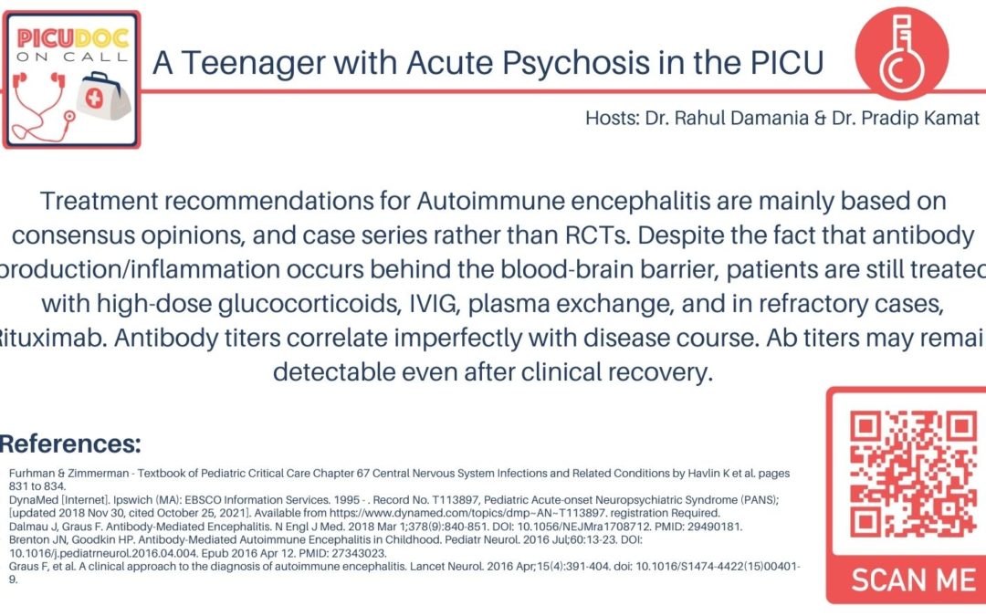
💡 In summary: The treatment recommendations for Autoimmune encephalitis are mainly based on consensus opinions, case series rather than RCTs. Despite the fact that antibody production/inflammation occurs behind the blood-brain barrier, in practice, patients are still treated with high-dose glucocorticoids, IVIG, plasma exchange, and in refractory cases rituximab. Antibody titers correlate imperfectly with disease course. Ab titers may remain detectable even after clinical recovery.
This concludes our episode on Anti-NMDA encephalitis. We hope you found value in our short, case-based podcast. We welcome you to share your feedback, subscribe & place a review on our podcast! Please visit our website picudoconcall.org which showcases our episodes as well as our Doc on Call management cards. PICU Doc on Call is by myself Pradip Kamat and Dr. Rahul Damania. Stay tuned for our next episode! Thank you!
- References:
- Furhman & Zimmerman – Textbook of Pediatric Critical Care Chapter 67 Central Nervous System Infections and Related Conditions by Havlin K et al. pages 831 to 834.
- DynaMed [Internet]. Ipswich (MA): EBSCO Information Services. 1995 – . Record No. T113897, Pediatric Acute-onset Neuropsychiatric Syndrome (PANS); [updated 2018 Nov 30, cited October 25, 2021]. Available from https://www.dynamed.com/topics/dmp~AN~T113897. registration Required.
- Dalmau J, Graus F. Antibody-Mediated Encephalitis. N Engl J Med. 2018 Mar 1;378(9):840-851. DOI: 10.1056/NEJMra1708712. PMID: 29490181.
- Brenton JN, Goodkin HP. Antibody-Mediated Autoimmune Encephalitis in Childhood. Pediatr Neurol. 2016 Jul;60:13-23. DOI: 10.1016/j.pediatrneurol.2016.04.004. Epub 2016 Apr 12. PMID: 27343023.
- Graus F, et al. A clinical approach to the diagnosis of autoimmune encephalitis. Lancet Neurol. 2016 Apr;15(4):391-404. doi: 10.1016/S1474-4422(15)00401-9.

