Welcome to PICU Doc On Call, A Podcast Dedicated to Current and Aspiring Intensivists. I’m Pradip Kamat and I’m Rahul Damania. We are coming to you from Children’s Healthcare of Atlanta – Emory University School of Medicine.
Welcome to our Episode with a 15 year old Male having hypotension and bradycardia.
Here’s the case presented by Rahul:
A 15 year old M presents to the PICU after sustaining an acute trauma. The patient was brought to the ER by his family after being on a boat and lifting a heavy object. He did not fall, sustain any head or extremity trauma, but did feel an achy non-radiating back pain shortly after the event. His grandmother states that the patient kept complaining about the back-pain and over the next few hours the patient became increasingly fatigued and flushed in the face. The patient was able to move his arms and legs and still walk, however family became concerned when the patient had abdominal fullness and was unable to urinate properly. He presents to the emergency department for further evaluation. In the emergency department he is noted to be awake however intermittently sleepy. His vital signs are notable for a HR of 58 bpm and a blood pressure of 85/60. He has 3/5 motor strength in his lower extremities with decreased sensation in his feet. Patellar reflexes are 1+ bilaterally. Rectal tone is normal. Acute resuscitation is begun for this patient.
To summarize key elements from this case, this patient has:
- Acute trigger
- Back pain
- Vital sign instability and lower motor neuron signs.
- All of which bring up a concern for a spinal cord injury.
- Let’s transition and discuss some history and physical exam components of this presentation:
- What are key history features in a child who presents with hypotension and bradycardia?
- As our worry is primarily spinal cord in etiology you would want to ask about trauma — this could be blunt or penetrating trauma
- You also would like to ask about the nature of the injury and scene. It is especially important to inquire with the pre-hospital providers about the nature of the injury and the patient course in transport. Besides our normal ABCs, it is important to ask the care taken regarding spinal cord restriction (such as use of a cervical collar or backboard)
- Another high yield history component when you think about hypotension and bradycardia is to assess for Numbness, weakness, or changes in bowel or bladder habits. In this case the patient had abdominal fullness which maybe due to bladder dysfunction.
This is a great summary of key history findings for patients who present with hypotension and bradycardia as it relates to spinal cord issues. Remember that patients who have Down’s syndrome may have a predilection to have lax ligaments especially in the upper verterbrae. As a result, you should have an increased index of suspicion if a Down’s Syndrome patient presents with hypotension and bradycardia in the presence or absence of trauma. In a study published in 2017 in Neurocrit Care it was estimated that about 20% of patients with Trisomy 21 may have atlantoaxial instability.
A great point which you just highlighted. Remember that when you approach hypotension and bradycardia, it is also important to focus on cardiac etiologies:
Bradycardia directly pulls down the cardiac output, potentially causing shock, and especially if you have a blunted vasoconstrictor response you can couple this bradycardia with hypotension.I do not want to delve too much out of the scope of today’s episode but there is a wide differential for bradycardia but specifically related to history you should consider intoxication as a cause of bradycardia and hypotension.
- This includes:
- Beta-blocker or calcium-channel blocker.
- Central alpha-2 agonist (e.g., clonidine, dexmedetomidine, guanfacine).
Going back to our case, are there some red-flag symptoms or physical exam components which you could highlight when you approach?
Yes, in this patient who we suspect spinal cord injury, we would like to perform a comprehensive neurological exam:
- Motor strength should be tested especially in the lower extremities
- Key muscle groups should be tested to determine level of injury
- Knee extensors are at L3
- Whereas your triceps and biceps can be assessed C5-C7.
On physical exam, this patient had a flushed face, and this could be related to an Interruption of sympathetic chain causing a horner’s syndrome like presentation.
Recall that Horner’s Syndrome is a triad of ptosis, miosis, and anhidrosis which can present as facial flushing.
During this spinal cord assessment it is important to perform a rectal exam to check for perianal sensation and rectal tone
- If at least 1 is normal in the acute setting, this suggests a sacral-sparing injury and thus an incomplete injury with the potential for some motor recovery
Other physical exam components includes assessing for priapism in male patients. Priapism in male patients may be present from abrupt loss of sympathetic tone to pelvic vasculature, causing a high-flow arterial priapism.
This is a great review of history & physical components for hypotension and bradycardia as a presentation of spinal cord injury — I think the key point here is to remember that this presentation is related to a loss of sympathetics and thus unopposed vagal tone which leads to the acute symptamology of Distributive shock with hypotension and bradycardia
To continue with our case, the patients labs were consistent with:
- Blood gas consistent with a metabolic acidosis
- A lactic acid of 4.6 mg/dL
- His coagulation panel and basic metabolic panel was within normal limits
- EKG was notable for sinus bradycardia with no evidence of heart block.
I would also like listeners to note that in patient with high cervical spinal cord injuries, the presence of hypercarbia suggesting hypoventilation may prompt for the need for early intubation
What did the imaging show in this patient?
- After stabilization, our patient underwent CT showing an T2 spinal cord injury. There was an associated T5 vertebral fracture.
Interesting this may have been related to his boat trauma. Remember listeners, that CT is very sensitive for defining bone fractures in the spine. Because CT is more sensitive than plain films, patients who are suspected to have a spinal injury and have normal plain films should also undergo CT. CT also has advantages over plain films in assessing the patency of the spinal canal. CT also provides some assessment of the paravertebral soft tissues and perhaps of the spinal cord as well, but is inferior in that regard to MRI.
OK, to summarize, we have:
- A 15 yo M who presents after trauma with hypotension, bradycardia, facial flushing and bladder dysfunction. This brings up the concern for spinal or neurogenic shock, the topic of our discussion today.
- Let’s start with a short multiple choice question:
- After a MVA, a 16 yo M presents with a HR 50 and MAP 45. Patient is obtunded, gurgling, and resuscitation efforts are begun. His hypotension does not improve with fluid resuscitation. A diagnosis of neurogenic shock is suspected. Stimulation of which of the following receptors is most likely to benefit this patient acutely?
- nicotinic ach receptors
- muscarinic ach receptors
- vasopressin -2 receptors
- alpha-1 receptors.
The correct answer is D. alpha-1 receptors. Remember that patients with neurogenic shock are devoid of sympathetics. Thus, you want to initiate sympathomimetics early. Some patients may require continuous infusion of norepinephrine, phyenlephrine, or dopamine.
As you think about our case, what would be your differential?
- First off I would make a distinction between Conus medullaris syndrome & Cauda Equina Syndrome.
- To start, the Conus medullaris is the terminal end of the spinal cord. If damaged, these children will have UMN weakness.
- They make have impaired sphincter control early, and Disturbances in urination
- Older children may be able to communicate a feeling of saddle anesthesia.
Pradip, what about cauda eqina syndrome?
Great question. So the Cauda equina is the lumbar and sacral roots caudal from the conus medullaris. These patients are going to have multiple nerves affected and may also have progressive incontinence.
In fact, studies have shown that Finding of urinary retention (post void residual > 100-200 mL) has 90% sensitivity for cauda equina syndrome.
A key distinction between the two is that cauda equaina syndrome in general has an asymmetric weakness with primarily LMN signs. These patient are going to have urinary retention that presents later from the onset of injury.
OK, to summarize, Conus medullaris syndrome you damage spinal cord, think early onset issues of bowel and bladder with UMN vs CE syndrome you have more damage of peripheral nerve roots and you in general will have a progressive inconitence with UMN signs.
RAHUL, I have also heard of this acronym, SCIWORA. What is this clinical entity?
SCIWORA stands for Spinal Cord Injury WithOut Radiographic Abnormality (SCIWORA)
In the pediatric population this differential is greater concern in pediatric population due to laxity of ligaments and weaker muscles
In this disorder, there is No discernible fracture on conventional films or computed tomography scans however patients may have spinal cord injury or on exam neurological deficits. The Mechanism is transient subluxation, stretching, or vascular compromise.
Finally, let’s contrast neurogenic shock with spinal shock — this is a subtle distinction clinically but has been described in the literature Rahul can you shed some light on that?
- Spinal Shock Syndrome with a temporary loss of neurologic function and tone below a level of an acute lesion
- Presents as flaccid paralysis, loss of sensation, loss of deep tendon reflexes, and urinary bladder incontinence
- Spinal reflexes often return in a predictive manner with the reflexes in the genital region among the first to reappear
- Spinal shock, when accompanied by hemodynamic compromise with loss of vasomotor tone, is generally going to be known as neurogenic shock. Neurogenic shock typically occurs in patients with a T5 injury and above however can be seen in any lesion throughout the spinal cord.
If our history, physical, and diagnostic investigation led us to neurogenic shock related to acute traumatic spinal cord injury as our diagnosis, what would be your general management of framework?
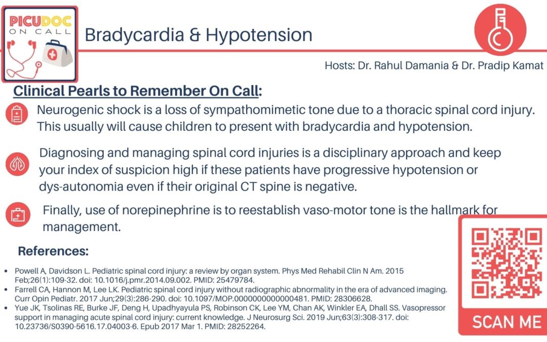
- We have made a key theme today regarding the interruption of autonomic pathways in the spinal cord causing decreased vascular resistance and bradycardia. As such, your management should be focused on resuscitation and re-initiation of sympathetic tone in the form of vasopressors.
- Remember that Patients with traumatic spinal cord injury may also suffer from hemodynamic shock related to blood loss and other complications.
- An adequate blood pressure is believed to be critical in maintaining adequate perfusion to the injured spinal cord and thereby limiting secondary ischemic injury.
- Bradycardia caused by cervical spinal cord or high thoracic spinal cord disruption may require external pacing or administration of atropine. However in studies atropine has not been shown to completely reverse neurogenic shock.
What about steroid use in spinal cord injuries?
- Methylprednisolone is the only treatment that has been suggested in clinical trials to improve neurologic outcomes in patients with acute, nonpenetrating TSCI. However, the evidence is limited, and its use is debated.
- In animal experiments, administration of glucocorticoids after a spinal cord injury reduces edema, prevents intracellular potassium depletion, and improves neurologic recovery – this is especially true within the first eight hours after injury.
- In 2013, based upon the available evidence, the American Association of Neurological Surgeons and Congress of Neurological Surgeons stated that the use of glucocorticoids in acute spinal cord injury is not recommended. Use of glucocorticoids in this setting appears to be declining.
- Let’s focus our management on the vasopressor use — as mentioned prior, vasopressors should be considered in cases of neurogenic shock esp if there is failure to respond to crystalloid, and no alternative diagnosis for hypotension.
- Your go to agents are going to be those that have a-lpha 1 activity to reestabllish vasomotor tone:
- Norepinephrine or Phenylephrine are your medications of choice in this setting
- Note phenylephrine may cause reflex bradycardia as this is a pure alpha one agonist.
In terms of prognosis:
- Adult studies have cited: 10%-20% of patients with spinal cord injuries do not survive to hospitalization.
- Most recovery starts within the first few weeks and plateaus in the first 3-6 months
- Better prognosis for ambulation include
- Younger age, decreased severity of impairment, incomplete injury, and lower level of injury
This is a great time for us to highlight the multi-disciplinary effort that goes into caring for these children. It is important in the acute setting to work closely with neurosurgery, ortho, neurology, and the critical care team and further in the subacute setting involving the rehabilitation team.
- Leading causes of death in children with spinal cord injury are respiratory conditions and pnuemonia so working closely with speech therapy for oromotor function is imperative in management.
- I would advise trainees and anyone interested to consider reading chapter 34 entitled shock states in Fuhrman & Zimmerman – Textbook of Pediatric Critical Care to review the hemodynamic patterns seen in our discussion of neurogenic shock.
This concludes our episode on Neurogenic shock. We hope you found value in our short, case-based podcast. We welcome you to share your feedback, subscribe & place a review on our podcast! Please visit our website picudoconcall.org which showcases our episodes as well as our Doc on Call management cards. PICU Doc on Call is hosted by myself Pradip Kamat and my cohost Dr. Rahul Damania. Stay tuned for our next episode! Thank you!
References:
Powell A, Davidson L. Pediatric spinal cord injury: a review by organ system. Phys Med Rehabil Clin N Am. 2015 Feb;26(1):109-32. doi: 10.1016/j.pmr.2014.09.002. PMID: 25479784.
Farrell CA, Hannon M, Lee LK. Pediatric spinal cord injury without radiographic abnormality in the era of advanced imaging. Curr Opin Pediatr. 2017 Jun;29(3):286-290. doi: 10.1097/MOP.0000000000000481. PMID: 28306628.
Yue JK, Tsolinas RE, Burke JF, Deng H, Upadhyayula PS, Robinson CK, Lee YM, Chan AK, Winkler EA, Dhall SS. Vasopressor support in managing acute spinal cord injury: current knowledge. J Neurosurg Sci. 2019 Jun;63(3):308-317. doi: 10.23736/S0390-5616.17.04003-6. Epub 2017 Mar 1. PMID: 28252264.

